It often occurs after motor vehicle accidents, beatings or falls. The impact from the front pushes the structures in the orbit posteriorly, causing an explosive fracture. In direct trauma to the orbit, fractures of the thin orbital floor adjacent to the maxillary sinus often occur, and rarely, fractures of other orbital walls are called blow-out fractures. The periorbital fat tissue may be displaced into the maxillary sinus through the defect in the orbital floor (enophthalmos). Extraocular muscles that move the eye may also be compressed in the fracture lines, eye movements may be restricted, and diplopia may occur in some gaze directions.Symptoms and signs: Due to the passage of the contents of the eye socket from the fracture site to the sinus, the eye is displaced inward or inward-downward, sometimes downward. In the first days, this depression of the eye may not be evident due to bleeding. Eye movement disorder and/or double vision may occur due to compression of extraocular muscles. Lacerations and ecchymosis may be seen around the eye. In any kind of orbital injury, the patient should be carefully examined and evaluated in terms of eyeball rupture, injury, and injury of other soft tissues in the eye socket. Indeed, the probability of eye injury accompanying orbital fractures varies between 10% and 25% in general.
The diagnosis is based on examination findings, computed tomography (CT) and MRI findings. It should also be noted that adjacent organs such as the nose, brain base and sinuses may also be injured and therefore consultation may be required.
Edema and hemorrhage caused by trauma should be surgically corrected in the early period after the edema and hemorrhage have passed. Enophthalmos, diplopia, infraorbital nerve hypoesthesia or anesthesia are indications for surgery. If there is a large fracture gap, it is recommended to repair it. Autogenous bone and cartilage grafts, titanium, polyethylene, hydroxyapatite, methylmethacrylate, ceramic, alloplastic materials can be used. If the defect is smaller than 10 mm, open reduction and fixation with a miniplate can be performed. Below, you can see the preoperative and postoperative pictures and radiologic images of a patient with such a trauma:
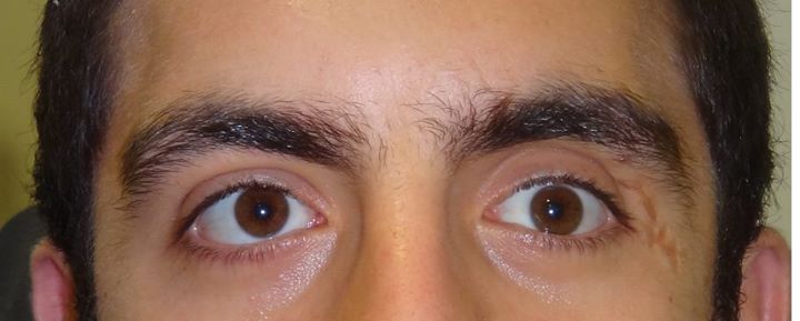
Figure 1: The left eye has shifted downwards after a soccer match.
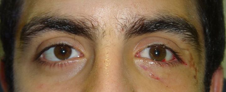
Figure 2: Condition of the left eye after surgery.
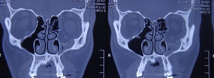
Figure 3: Preoperative radiologic view. The base of the left eye socket is fractured, the eye and surrounding structures have shifted downward, and the maxillary sinus is filled.
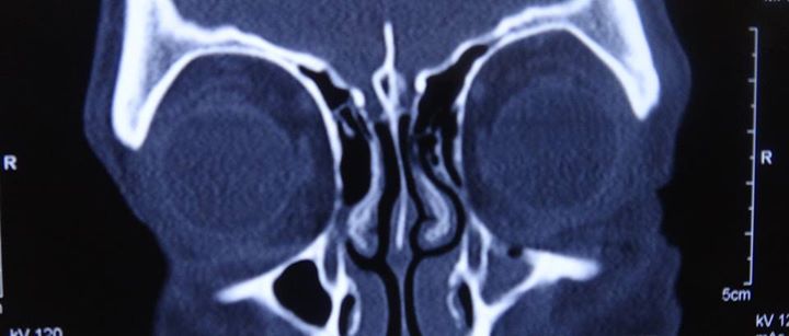
Figure 4: Postoperatively, the left eyeball and surrounding tissues are intact and the base fracture is closed with the material placed on the base of the orbit.